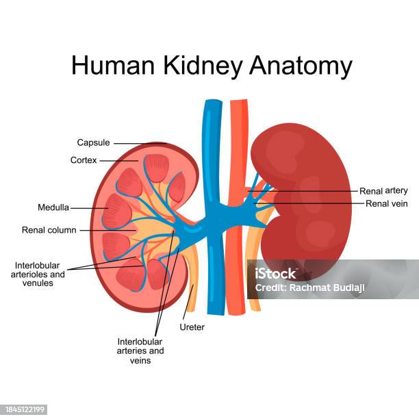
0514 Anatomy Of Kidney Medical Images For PowerPoint 1 Biology Diagrams Learn about the structure and function of the kidneys, the body's filtration system. See diagrams and images of the kidneys and their parts, and find out about common diseases and conditions that affect them. Learn about the kidneys, their structure, function, and blood supply with labeled diagrams. Explore the external and internal anatomy of the kidney, including the renal capsule, cortex, medulla, pyramid, nephron, and more. Learn about the external and internal anatomy of the kidney, including its location, blood supply, nephrons, and urine formation. See diagrams, videos, and review questions to test your knowledge.

Learn about the kidneys, their function, location, internal structure and blood supply. See diagrams, 3D models and prosections of the kidneys and their coverings, pyramids, calyces and pelvis.
Biology LibreTexts Biology Diagrams
Learn about the structure, function, and location of the kidneys, the bean-shaped organs that filter blood and excrete waste. See diagrams and illustrations of the kidney anatomy, nephrons, and urinary system. This Osmosis High-Yield Note provides an overview of Anatomy and Physiology of the Renal System essentials. All Osmosis Notes are clearly laid-out and contain striking images, tables, and diagrams to help visual learners understand complex topics quickly and efficiently. Find more information about Anatomy and Physiology of the Renal System:
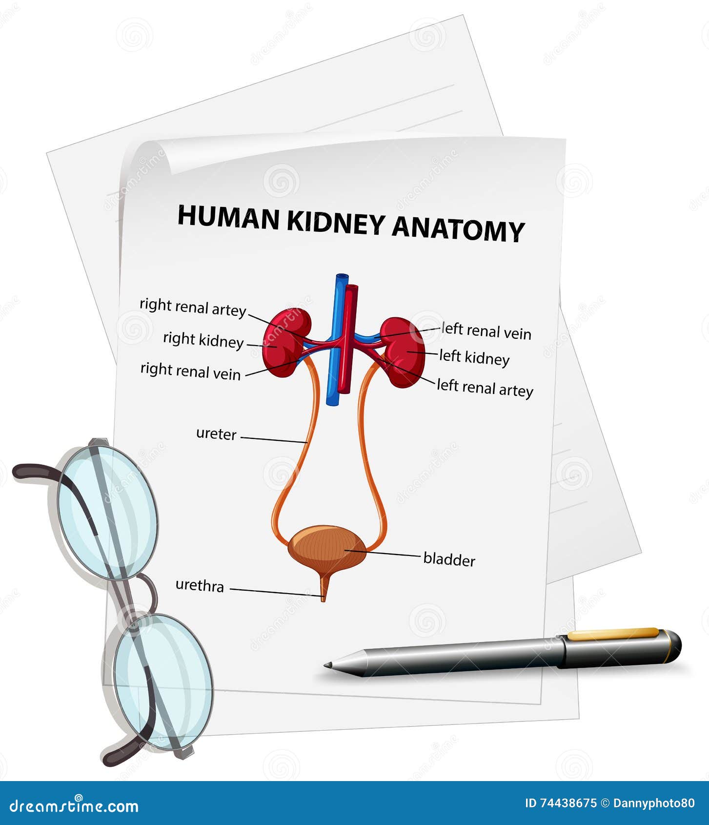
Learn about the kidneys, their functions, and their anatomy with diagrams and quizzes. Find out how they regulate blood pressure, acid-base balance, hormones, and more. Kidney Anatomy. The shape of each kidney gives it a convex side and a concave side. You can see this clearly in the detailed diagram of kidney anatomy shown in Figure \(\PageIndex{3}\). The concave side is where the renal artery enters the kidney and the renal vein and ureter leave the kidney. This area of the kidney is called the hilum.